
Moreover, these plugins require very high temporal resolution for tracking individual blebs. Due to their unique shape, leader blebs are not likely to be accurately identified by this method. Other plugins for analyzing blebs rely on a characteristic change in curvature. Blebs can form and retract in less than a minute, whereas cells displaying LBBM migrate at a speed of about 30 μm per hour. Bleb dynamics are fast compared to the speed of migration. The study of LBBM presents a quantitative challenge. Thus, LBBM is an important new mode of migration that cells may adopt under confinement. The hallmarks of LBBM have also been observed in vivo, including within developing Zebrafish embryos and tumors by intravital imaging. Accordingly, as cells move in the direction of this bleb, it has been termed a ‘leader bleb’.

Importantly, with non-specific friction, it is this cortical actin flow that provides the motive force for cell movement. Within these stable blebs, cortical actin flows from the bleb tip to the neck, which is enriched with myosin and separates the bleb from the cell body. However, when cells are confined down to 3 μm (which has been found to be optimal) they may form a very large and stable bleb. In tissue culture, blebs are normally dynamic–forming and retracting in less than a minute. Accordingly, cells with elevated intracellular pressure often display numerous blebs. Blebs form in response to a local separation between the PM and underlying cortical actin cytoskeleton. Ī hallmark of amoeboid migration is the formation of Plasma Membrane (PM) blebs. Under conditions of high (mechanical) confinement, a diverse range of cell types have recently been shown to undergo a phenotypic transition to fast amoeboid or Leader Bleb-Based Migration (LBBM). Under conditions that more closely resemble the tissue environment, cells have been shown to rapidly switch between a growing list of migration modes, optimizing their migratory potential based on their immediate (tissue) environment. A large portion of cell migration research has been conducted on glass substrates, which favors (integrin-dependent) mesenchymal motility. Accordingly, the de-regulation of cell migration is a hallmark of disease. The funders had no role in study design, data collection and analysis, decision to publish, or preparation of the manuscript.Ĭompeting interests: The authors have declared that no competing interests exist.Ĭell migration mediates diverse physiological processes, including embryonic development, immune surveillance, and wound healing.

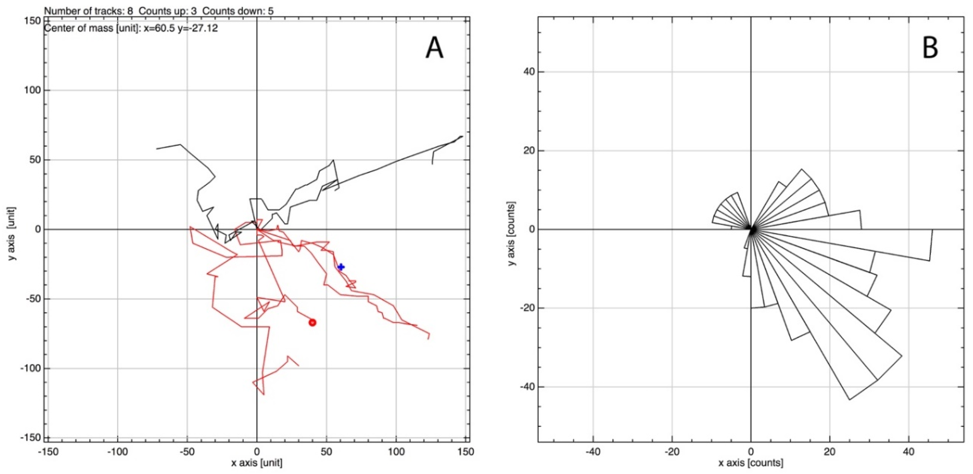
688232) (DOI: ), and a Cancer Research Scholar Grant from the American Cancer Society (ACS award no. This is an open access article distributed under the terms of the Creative Commons Attribution License, which permits unrestricted use, distribution, and reproduction in any medium, provided the original author and source are credited.ĭata Availability: All relevant data are within the manuscript and its Supporting Information files.įunding: This work was supported by start-up funds from the Albany Medical College, a Young Investigator Award from the Melanoma Research Alliance (MRA award no. Received: DecemAccepted: ApPublished: April 29, 2022Ĭopyright: © 2022 Vosatka et al. PLoS ONE 17(4):Įditor: Shengyu Yang, Penn State College of Medicine, UNITED STATES Furthermore, that F-tractin increases cell stiffness, which was found to correlate with a decrease in migration, thus reaffirming the importance of cell mechanics as a determinant of Leader Bleb-Based Migration (LBBM).Ĭitation: Vosatka KW, Lavenus SB, Logue JS (2022) A novel Fiji/ImageJ plugin for the rapid analysis of blebbing cells. We also demonstrate, using flow cytometry, that live markers increase total levels of F-actin. As validation, we show that Analyze_Blebs can detect significant differences in cell migration and morphometrics, such as the largest bleb size, upon introducing different live markers of F-actin, including F-tractin and LifeAct tagged with green and red fluorescent proteins. Here, we demonstrate that a novel Fiji/ImageJ-based plugin, Analyze_Blebs, can be used to quickly obtain cell migration parameters and morphometrics from time lapse images.
However, as this is a nascent area of research, few tools are available for the rapid analysis of cell behavior. When confined, cells have recently been shown to undergo a phenotypic switch to what has been termed, fast amoeboid (leader bleb-based) migration.


 0 kommentar(er)
